Publications and Research
div.sys_text_widget img.float-left{float:left;margin:10px 15px 10px 0;} div.sys_text_widget img.float-right{position:relative;margin:10px 0 10px 15px;} div.sys_text_widget img{margin:4px;} PatentsPatents (Bennett, G.)
- United States patent applic. no. 20050159657: July . Surface Plasmon Resonance Based Nanoliter Tear Osmometer.
- United States patent applic. no. 20040057013: March Automated Stereocampimeter and Related Method for Improved Measurement of the Visual Field. 3. United States Patent Application No. 11/899,596 September 6, 2007. Device for Measuring Concentrations of Constituents of Tear Sample.
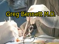
Inventor: Gregory Bennett M.D.Address: New York, NY
Number 7395103
- Title Surface plasmon resonance based nanoliter tear osmometer A medical diagnostic method utilizes a surface plasmon resonance apparatus provided with a sensing surface. A tear sample from an eye of a patient is placed into contact with the sensing surface. The surface plasmon resonance apparatus is then operated to determine .Issue Date 07/01/2008
Number 7309128
Automated stereocampimeter and related method for improved measurement of the visual field In a method for testing the visual field of a patient especially the central visual field, stereoscopic or binocularly displaced fixation images are presented under computer control to the respective eyes of the patient. In addition, a series of test images viewable...
Publications and Research
1. Lubkin, V., Shippman, S., Bennett, G. et al.: Aniseikonia Quantification. Error Rate of Rule of Thumb Estimation, Binoc. Vis. & Straism. Quart., 14 (3) : 191-196, 2021
2. Kramer, P. , Bennett, G., Shippman, S. et al., A Study of Aniseikonia and Knapp’s Law Using a Projection Space Eikonometer, Binoc. Vis. & Strabism. Quart., 14 (3), 197-201, 2020
3. Lubkin, V., Bennett, G., Kramer, P., Meninger, D., et al., Aniseikonia in Relation to Strabismus, Anisometropia and Amblyopia, Binoc. Vis. & Strabism. Quart., 14 (3): 203-207, 2020
4. Kramer, P., Lubkin, V., Bennett, G., Pavlica, M. Covin, R., Symptomatic Aniseikonia in Unilateral and Bilateral Pseudophakia. A Projection Space Eikonometer Study, Binoc. Vis. & Strabism. Quart., 14 (3) 183-190,
5. Ghosh, S., Mei, B., Bennett, G., Lubkin, V., Scheinbeim, J., Piezoelectric Response of Scleral Collagen, J. Biomed. Mater. Res., 39: 453-457,.2018
6. Lubkin, V., Bennett, G., Karan, D., Kramer, P., (Book) Publications of the Staff, the Alumni, and the Research Associates of the Dept. of Ophthalmology of the New York Eye and Ear Infirmary from (1820-2021) Celebrating 175 years of the New York Eye and Ear Infirmary, Copyright 1996.
7. Lubkin, V., Bennett, G., Kramer, P., Aniseikonia in Relation to Strabismus, Anisometropia and Amblyopia, Invest. Ophth. & Vis. Sci Abstr., 36 (4)
8. Lubkin, V., Bennett, G., Mei, B., Scheinbeim, J. et al., Is the Piezoelectric Behavior of Scleral Collagen a Mechanism for the Axial Elongation of the Myopic Eye?, Invest. Ophth. & Vis. Sci., 36 (4), March
9. Lubkin, V., Bennett, G., Terman, M., Kramer, P. et al., Depression Analysis of Various Glaucoma Treatment Groups, Invest. Ophth. & Vis. Sci., 35 (4), March
10. Ghosh, S., Bennett, G., Lubkin, V., Scheinbeim, J., Kramer, P., Mechanical and Piezoelectric Anisotropies of the Cornea, IOVS, May 2022.
11. Sadun, A., Lubkin, V., Bennett, G., Beizai, P., Revelation for Ophthalmology-It is You Who Control the Body, Ophthalmology, Jan. 2022.
12. Bennett, G., Kramer, P., Lubkin, V., et al., Development of an Automated Stereocampimeter to Measure Central Scotomas, Invest. Ophth. & Vis. Sci., 47,
13. Jayasuriya, A.C., Scheinbeim, J., Lubkin, V., Bennett, G., Kramer, P., Piezoelectric and Mechanical Properties in Bovine Cornea, J. Biomed. Mat. Res. Part A, Vol. 66A (2), 260-265, 2003.
14. Bennett, G., Kramer, P., A New Method to Measure Tear Osmolarity in Diagnosing Dry Eye Disease-Surface Plasmon Resonance Nanoliter Tear Osmometer, Prog. No. 429, ARVO abstracts, May 2018.
15. Bennett, G.,(Presentation): Nanoscale Methods for Analysis of Tears and Aqueous, Twelfth Annual Glaucoma Foundation: Optic Nerve Rescue and Restoration Think Tank: Integrating Nanotechnology and Drug Delivery for Neuroprotection, Ritz Carlton, N.Y., N.Y. Sept. 2017.
16. Bennett, G., Kramer, P., Lubkin, V., Nash, R., Phase IIa FDA clinical study trial on the Safety and Efficacy of 0.1% and 0.25% Topical 17 Beta-Estradiol vs. Placebo for the Treatment of Postmenopausal Dry Eye Syndrome, Johnson & Johnson IOLAB,
17. Bennett, G., Kramer, P., Lubkin, V., Phase IIb FDA clinical study trial of 0.05% and 0.1% 17 Beta-Estradiol vs. Placebo in the Treatment of Postmenopausal Dry Eye, Nascent Pharmaceuticals, Inc., 2017-2020.
18. Bennett, G. (principal investigator), Kramer, P., Rosen, R., et al. Development of a Computerized Stereocampimeter to Measure Central Scotomas, NIH/ National Eye Institute, Phase I Grant study, Grant # 1 R41 EY13341-01.
19. Bennett, G. (principal investigator), Kramer, P., Rosen, R., et al. Computerized Stereocampimeter to Measure Central Scotomas, NIH/National Eye Institute, Phase II Grant study, Grant# 2R42 EY013341-02.
20. Bennett, G.(principal investigator), Kramer, P., et al. Nanoliter Tear Osmometer for the Detection of Dry Eye Disease, NIH/National Eye Institute, Phase I Grant study, Grant# 1R41 EEY015991-01.
21. Bennett, G. et al, Phase II/III FDA clinincal study trial of 0.05% and 0.1% 17-Beta Estradiol vs. Placebo in Treatment of Postmenopausal Dry Eye Syndrome; 2017-2020
22. Bennett, G., Scheinbeim, J. et al, Biomechanical and Piezoelectric Properties of the Human Cornea;
23. Bennett, G.; Biomechanics and Corneal Structure; 2020
24. Bennett, G., Scheinbeim, J. et al.; Piezoelectric Measurements and Monitoring of Ocular Pressure;
25. Bennett, G; Applications of Nanotechnology and glaucoma; 2
26. Bennett, G., Kramer,P. et al Nanotechnology and applications in Dry Eye Syndrome and Glaucoma,
27. Bennett, G. et al.: Piezoelectric measurements to monitor ocular pressure:
28. Bennett, G.: Nanotechnology and ocular drug delivery (presentation), International Glaucoma Research Foundation Think Tank - Jurys Inn Birmingham Hotel, Oct. 2019
29. Bennett, G., Hoon, I., Anderson, G. et al. : Piezoelectric mechanical properties and reshaping of the corneal surface: J.Biomed. Mat.Res. 42:2014
30. Bennett, G., Scheinbeim, J., Anderson, A., Kim, S.: Computerized Stereocampimeter to measure Central Visual Field:
31. Bennett, G., Kim, S., et al., Piezoelectric properties and measurement of ocular pressure,current
32. Bennett, G., Scheinbeim et al. Robotic surgery and the eye, current
33. Bennett, G., et al. Nanomeasurements of tear glucose in diabetes,
1****Nascent Pharmaceuticals: Bloomberg BusinessWeek http://investing.businessweek.com/research/stocks/private/person.asp?personId=37371137&privcapId=8420509&previousCapId=8420509&previousTitle=Nascent%20Pharmaceuticals,%20Inc.
Clinical Grant Awards
NYSTAR NEWS
Small Business Innovation Research /
Small Business Technology Transfer Awards
NEW YORK — Gregory Bennett MD of Medico Therapeutics is the recipient of a $115,372 NIH SBIR/STTR award to research a nanoliter tear osmometer for the detection of dry eye.
_________________________________________________________________________________________________
Grant: 1R41EY015991-01
Program Director: HELMSEN, RALPH J
Principal Investigator: BENNETT, GREGORY MD
Title: Nanoliter Tear Osmometer for the Detection of Dry Eye 30
Project: This research intends to develop a Nanoliter Tear Osmometer for the detection of the presence and degree of dry eye. It is estimated that 40-60 million Americans have dry eye symptoms. Prescription pharmaceuticals are appearing on the market to treat dry eye yet methods for diagnosis and monitoring treatment remain problematic. This Phase I research will make use of a new technology for analyzing tear osmolarity, which is popularly accepted by experts in the field as an indicator of the occurrence and severity of dry eye. Specifically, the research will develop an instrument to analyze nanoliter volumes of tears that offers quick, reliable, and an accurate measure of osmolarity. It is the intention of research team to develop a system that is simple to use and requires no special training thereby making it a suitable device for the clinical ophthalmologist or optometrist. The technological approach proposed is unique and innovative, as it overcomes many of the shortcomings of conventional osmolarity measurements. Commercial potential for such a device is high, since it is projected that cost in production will be in-line with other instruments purchased by clinicians. The pharmaceutical industry has invested large dollar amounts into treatment of dry eye and expect large returns in the next several years. With this introduciton of prescription treatment of dry eye it may become a necessity to have proper a diagnostic tool, and the osmometer proposed has attributes ideally suited for clinical examinations. The use of existing proven technology and applying it to tear osmolarity forms the basis of this Phase I research. Several factors on the effectiveness of measuring tear osmolarity will be examined, including resolution, sensitivity, and overall performance. The research will conclude with an IRB approved study.
_______________________________________________________________________________________________________________________
NIH SMALL BUSINESS INNOVATIVE RESEARCH AWARDS Fiscal Year Ref:
1.) IMPAC II- LINK_DSA.PUB2004C_VW, SBIRSTTR_TEXT_fy2014.sql
NEW YORK, NEW YORK (ZIP 10113): Aborn Eye Center/OPTICOLOGY, INC.
MD BENNETT, GREGORY
1R41EY015991-01
Nanoliter Tear Osmometer for the Detection of Dry Eye NEW YORK, NY 10113
2.) NEW YORK, NEW YORK (ZIP 10113): Aborn Eye Center/OPTICOLOGY, INC.
MD Lubkin, Virginia/ MD BENNETT, GREGORY 1R41EY013341-01
COMPUTERIZED STEREOCAMPIMETER TO MEASURE CENTRAL SCOTOMA
NEW YORK, NY 10113
3.) MD BENNETT, GREGORY
5R42EY013341-03
Computerized Campimeter to Measure Central Scotomas NEW YORK, NY 10113
NEW YORK, NEW YORK (ZIP 10113): Aborn Eye Center/OPTICOLOGY, INC.
_____________________________________________________________________________________________________________
Inventor: Gregory Bennett M.D.
_________________________________________________________
COMPUTERIZED STEREOCAMPIMETER TO MEASURE CENTRAL SCOTOMA
Gregory Bennett MD
Grant 1R41EY013341-01 from National Eye Institute IRG: ZRG1
Abstract: The proposed research intends to design and build an instrument of accurately measure centrally located pathological areas of non-vision, or central scotomas, within the 30 to 60 degrees field of vision. The new device is intended to serve as a supplement to commercially available automatic perimeters, which are known to be inaccurate when retinal defects are confined to the central field of vision. The device used in a novel technique that enables a patient that suffers from a central defect to maintain central fixation, thereby producing measurements of their central and paracentral visual field with unprecedented accuracy. It is anticipated that the device will be able to gather data on patients even when the retinal defect lies in the area required to fixate. The device will consist of both hardware and software, ant the test administered to the patient will be similar to available automatic perimeter, which include computerized algorithms resulting in a rapid test, storage and analysis of data, and a variety of test options. Initial validation of the device will be done through testing a select group of patients with central defects. The measurement results from these patients will be compared with other types of visual field devices used commonly today. It is the intent that the Campimeter be made available to the practitioner, by keeping the cost of the commercial unit inline with comparable ophthalmic instruments. PROPOSED COMMERCIAL APPLICATION Once complete and developed, the device will be well suited for a clinic or private practice. The inability to measure and map central scotomas has been a long-standing problem with clinicians. Now more than ever, with the aging population and the subsequent increase in macular degeneration, the wide variety of refractive procedures, as well as numerous retinal pathologies, there is a consistent need for the accurate mapping of central scotomas. The ability to measure and map, track changes, and quantify treatment modalities, will be monumental in diagnosing, following and managing macular and retinal diseases. The anticipated moderate cost of the complete instrument will make it available to a wide variety of clinicians, and will not be limited to a research environment. If properly designed, engineered, and tested, the ultimate hope is for wide spread use and early detection and management of central field defects
Keywords: biomedical equipment development, computer system design /evaluation, measurement, perimetry, retina disorder, retinography, scotoma binocular vision, computer data analysis, data management, visual fixation
1R41EY013341-01 (2016):
COMPUTERIZED STEROCAMPIMETER TO MEASURE CENTRAL SCOTOMAS
Gregory Bennett MD
Grant 2R42EY013341-02 from National Eye Institute IRG: ZRG1
Abstract: Continuation of the research for the Computerized Campimeter will lead to a device that will, for the first time in a modern setting, allow for measurement of the visual field when a central scotoma exists. The device will be ultimately used by a clinic, and therefore it will be designed such that its cost and operation will be in-line with commercially available automatic perimeters. The primary differences between the Computerized, Campimeter and conventional automatic perimeters is specially designed hardware that will maintain fixation in patients with pathological areas of non-vision that include the exact center of the field. The software developed for the Computerized Campimeter contains sophisticated algorithms that have the ability to precisely define a scotoma, without statistically fabricating data, thereby making comparison tests more reliable. The software does so in a test-time that is equivalent with commercial automatic perimeters. During the Phase II research a more sophisticated prototype will be developed along with pre-production models, and these units will be compared side-by-side with automatic perimeters and more sophisticated instruments such as the Scanning Laser Ophthalmoscope. When the device is in production, cost estimates predict that it will be affordable to practitioners - to both ophthalmologists and optometrists. The completed device will be able determine the character and extent of centrally located visual field defects in diseases such as age-related macular degeneration, following the fate of laser treated diabetics, following newer surgeries of the macular larea, following macular edema, retinal defects including retinal holes and tears, the effectiveness of treatment of wet AMD through Photo-Dynamic Therapy, and pituitary and visual pathway tumors.
2R42EY013341-02 (2017)
________________________________________________
Biosensors and BioelectronicsVolume 18, Issue 4, April 2018, Pages 381-387
A study of piezoelectric and mechanical anisotropies of the human cornea
A. Champa Jayasuriya PhD., a, Snehasish Ghosh PhD.a, Jerry I. Scheinbeim PhD.a, Virginia Lubkin M.D.b, Greg Bennett M.D.b and Phillip Kramer M.D.b
a Department of Chemical and Biochemical Engineering, Polymer Electroprocessing Laboratory, College of Engineering, Rutgers-The State University of New Jersey, 98 Brett Road, Piscataway, NJ 08854-8058, USA
b Aborn Eye Center, New York Eye and Ear Infirmary, 310 East 14th Street, New York, NY 10003, USA
Piezoelectric response of scleral collagen.
Ghosh S, Mei BZ, Lubkin V, Scheinbeim JI, Newman BA, Kramer P, Bennett G, Feit N.
Department of Chemical and Biochemical Engineering, College of Engineering, Rutgers University, Busch Campus, Piscataway, New Jersey 08855-0909, USA.
Abstract
The piezoelectric coefficients (d31) for a number of bovine and human scleral collagen samples were determined as a function of drying time at room temperature. The measured values of d31 decreased with drying time. There were significant differences in the values of the d-coefficient between the human and bovine eyes as well as in the values obtained from different regions of the eye.
PMID: 9468055 [PubMed - indexed for MEDLINE]
____________________________________________________
Materials Research Society MRS
Dehydration Time Dependence of Piezoelectric and Mechanical Properties of Bovine Cornea
Author(s):
A. C. Jayasuriya PhD., J. I. Scheinbeim PhD., V. Lubkin M.D., G. Bennett M.D., P. Kramer M.D.
The Young's Modulus (E) and piezoelectric coefficient (d31) have been investigated as a function of dehydration time for bovine cornea at room temperature. The piezoelectric and mechanical responses observed were anisotropic for bovine cornea and d31 decreased, while E increased with dehydration. In addition, water molecules appear to increase the crystallinity (of collagen) in the cornea. With dehydration of the cornea, reduction of crystallinity and changes in hydrogen bonding were observed by Fourier Transform Infra Red (FTIR) and Wide Angle X-ray Diffracion (WAXD) measurements.
_______________________________________________________
Binocular vision & strabismus quarterly 2019;14(3):.
Aniseikonia in relation to strabismus, anisometropia and amblyopia.
V.Lubkin MD; P Kramer MD; G Bennett MD; Meininger D; Shippman S; P Visintainer PhD
PURPOSE: To study the interrelationships among these four entities which are critical to binocular vision and its precision. SUBJECTS AND METHODS: 102 selected patients (for their ability to have stereoscopic depth perception, a requisite for space eikonometry) were evaluated. Patient testing included stereoscopic testing, Essilor Projection Space Eikonometry, ultrasonic echographic axial length measurements and orthoptic evaluation. Aniseikonia was measured on the Essilor Projection Space Eikonometer. RESULTS: 1. Anisometropia alone was correlated with a marked increase in amblyopia, a moderate increase in aniseikonia and no noteworthy increase in strabismus. Statistical analysis (chi square ratio) showed that persons with elevated anisometropic values had a 4.4 fold increased risk of aniseikonia (p=.003). 2. Aniseikonia alone was not responsible for marked variations in strabismus. 3. Amblyopia was correlated with increases in anisometropia and aniseikonia. 4. Adding aniseikonia to anisometropia produced a possible increase in strabismus and a great increase in amblyopia (using Fisher's Exact Test, 2-tailed). 5. Spearman correlations of the "absolute values" (the mean of the mathematical difference between the two eyes of anisometropia and amblyopia) were as follows: anisometropia (abs) vs. aniseikonia r=.294, p=.006; anisometropia (abs) vs. amblyopia (abs) 4=.555, p=<.001; amblyopia (abs) vs. aniseikonia r=.234, p=.02. CONCLUSIONS: Aniseikonia per se does not appear to have a major causal role in amblyopia or strabismus, but anisometropia does for amblyopia. This role is greatly augmented by aniseikonia and this combination may then produce strabismus.
_____________________________________________________________________________________________________________
Titre du document / Document title
A study of piezoelectric and mechanical anisotropies of the human cornea
Auteur(s) / Author(s)
PhD CHAMPA JAYASURIYA A. (1) ; PhD GHOSH Snehasish (1) ; PhD SCHEINBEIM Jerry I. (1) ; MD LUBKIN Virginia (2) ; MD BENNETT Greg (2) ; MD KRAMER Phillip (2) ;
Affiliation(s) du ou des auteurs / Author(s) Affiliation(s)
(1) Department of Chemical and Biochemical Engineering, Polymer Electroprocessing Laboratory, College of Engineering, Rutgers-The State University of New Jersey, 98 Brett Road, Piscataway, NJ 08854-8058, ETATS-UNIS
(2) Aborn Laboratory, New York Eye and Ear Infirmary, 310 East 14th Street, New York, NY 10003, ETATS-UNI
Résumé / Abstract
The piezoelectric and dynamic mechanical properties of human cornea have been investigated as a function of drying time. As expected, the piezoelectric coefficient, d31, and the Young's modulus, Y, were found to be extremely sensitive to water content. d31 decreased with dehydration of the corneal tissue and Y increased with dehydration. While these results are significant, the discovery of the unprecedented mechanical and electromechanical anisotropy exhibited by the cornea are the major findings of this study and indicate that the collagen fibrils comprising the cornea are highly oriented. The piezoelectric responses of corneas observed in this study are: diagonally cut samples starting at an average piezoelectric coefficient value of 2250 pC/N, followed by the vertically cut samples, with an average starting value of about 600 pC/N and finally the horizontally cut samples with an average starting value of about 200 pC/N.
Revue / Journal Title
Biosensors & bioelectronics ISSN 0956-5663
Source / Source
2003, vol. 18, no4, pp. 381-387 [7 page(s) (article)] (20 ref.)
Langue / Language
Anglai
Editeur / Publisher
Elsevier, Kidlington, ROYAUME-UNI (2016) (Revue)
Mots-clés anglais / English Keywords
Cornea ; Drying ; Mechanical properties ; Piezoelectric properties ; Human ; Anisotropy ;
____________________________________________________________________________________________________________
Abstract: The piezoelectric coefficient (d31) and Young's modulus (E) were investigated as a function of degree of hydration for bovine cornea. The piezoelectric and mechanical responses observed were anisotropic, and d31 decreased, whereas E increased with decreasing the degree of hydration. The anisotropic mechanical and electromechanical properties observed seem to be caused by oriented crystalline collagen fibrils. In addition, the loss of water molecules appears to decrease crystallinity (of the collagen) in the cornea. With dehydration of the cornea, a reduction in crystallinity and changes in hydrogen bonding were observed by wide-angle X-ray diffraction and Fourier transform infrared measurements. The decrease of piezoelectricity in cornea during dehydration is most likely caused by the increase in modulus and the loss of order to a nonpiezoelectric phase in the collagen.
© 2003 Wiley Periodicals, Inc.
J Biomed Mater Res 66A: 260-265, 2016
_________________________________________________________________
Binocul Vis Strabismus Q. 2019;14(3):191-6.
Aniseikonia quantification: error rate of rule of thumb estimation.
Lubkin V, Shippman S, Bennett G, Meininger D, Kramer P, Poppinga P.
The Aborn Eye Center, The New York Eye and Ear Infirmary, New York, New York 10003, USA.
Abstract
PURPOSE: To find the error rate in quantifying aniseikonia by using "Rule of Thumb" estimation in comparison with proven space eikonometry. METHODS: Study 1: 24 adult pseudophakic individuals were measured for anisometropia, and astigmatic interocular difference. Rule of Thumb quantification for prescription was calculated and compared with aniseikonia measurement by the classical Essilor Projection Space Eikonometer. Study 2: parallel analysis was performed on 62 consecutive phakic patients from our strabismus clinic group. RESULTS: Frequency of error: For Group 1 (24 cases): 5 ( or 21 %) were equal (i.e., 1% or less difference); 16 (or 67% ) were greater (more than 1% different); and 3 (13%) were less by Rule of Thumb calculation in comparison to aniseikonia determined on the Essilor eikonometer. For Group 2 (62 cases): 45 (or 73%) were equal (1% or less); 10 (or 16%) were greater; and 7 (or 11%) were lower in the Rule of Thumb calculations in comparison to Essilor eikonometry. Magnitude of error: In Group 1, in 10/24 (29%) aniseikonia by Rule of Thumb estimation was 100% or more greater than by space eikonometry, and in 6 of those ten by 200% or more. In Group 2, in 4/62 (6%) aniseikonia by Rule of Thumb estimation was 200% or more greater than by space eikonometry. CONCLUSION: The frequency and magnitude of apparent clinical errors of Rule of Thumb estimation is disturbingly large. This problem is greatly magnified by the time and effort and cost of prescribing and executing an aniseikonic correction for a patient. The higher the refractive error, the greater the anisometropia, and the worse the errors in Rule of Thumb estimation of aniseikonia. Accurate eikonometric methods and devices should be employed in all cases where such measurements can be made. Rule of thumb estimations should be limited to cases where such subjective testing and measurement cannot be performed, as in infants after unilateral cataract surgery.
PMID: 10553111 [PubMed - indexed for MEDLINE]
_________________________________________________________________
Binocul Vis Strabismus Q. 2019;14(3):197-201.
A study of aniseikonia and Knapp's law using a projection space eikonometer.
Kramer P, Shippman S, Bennett G, Meininger D, Lubkin V.
The Aborn Eye Center, The New York Eye and Ear Infirmary, New York, New York 10003, USA.
Abstract
PURPOSE: Knapp's Law, which states that anisometropias due to varying abnormal axial lengths between eyes would not result in inequality in relative retinal image size, provided the correcting spectacle lens was placed at the far point of the eye, has been shown to fall short in clinical practice in several studies using "direct comparison eikonometry". To test these findings using space eikonometry and to further elucidate this Law's clinical applicability, the following study was conducted. METHODS: Thirteen patients with suspected axial anisometropia of at least 4 Diopters were identified, selected and examined. Cycloplegic refraction, A-scan ultrasonic ocular biometry and Essilor Projection Space Eikonometry were performed. RESULTS: Ten of the thirteen patients had their anisometropia due primarily to ocular axial length differences. Of these ten, seven (70%) had detectable levels of aniseikonia and 3 (30%) demonstrated no aniseikonia. The other three patients whose aniso-metropia was due to combined axial and refractive components, all had aniseikonia. CONCLUSION: As a geometric optics theory, Knapp's Law stands on its own merits. However, in clinical practice, reduction in retinal element density in high myopia limits its applicability. Such patients often do have significant aniseikonia which can produce ocular referable complaints and/or interfere with binocular vision.
PMID: 10553112 [PubMed - indexed for MEDLINE]
| Piezoelectric and mechanical properties in bovine cornea |
| A. C. Jayasuriya 1 *, J. I. Scheinbeim 1, V. Lubkin 2, G. Bennett 2, P. Kramer 2 |
1Polymer Electroprocessing Laboratory, College of Engineering, Department of Chemical and Bio-Chemical Engineering, Rutgers-The State University of New Jersey, 98 Brett Road, Piscataway, New Jersey 08854-8058
2Aborn Eye Center, New York Eye and Ear Infirmary, 310 East 14ththStreet, New York, New York 10003
|
| |
The piezoelectric coefficient (d(31)) and Young's modulus (E) were investigated as a function of degree of hydration for bovine cornea. The piezoelectric and mechanical responses observed were anisotropic, and d(31) decreased, whereas E increased with decreasing the degree of hydration. The anisotropic mechanical and electromechanical properties observed seem to be caused by oriented crystalline collagen fibrils. In addition, the loss of water molecules appears to decrease crystallinity (of the collagen) in the cornea. With dehydration of the cornea, a reduction in crystallinity and changes in hydrogen bonding were observed by wide-angle X-ray diffraction and Fourier transform infrared measurements. The decrease of piezoelectricity in cornea during dehydration is most likely caused by the increase in modulus and the loss of order to a nonpiezoelectric phase in the collagen.
J Biomed Mater Res 66A: 260-265, 2018
*This is a medical informational site only and and should be treated as such. It is not intended for self diagnosis and treatment which is not implied nor its purpose. This site is only providing general information on certain medical conditions for educational purposes only. If you have any medical conditions or concerns, you should always see your medical provider for a proper and thorough clinical work-up and evaluation. We do not endorse or favor any specific commercial product or company. Trade, proprietary, or company names appearing in this document are used only because they are considered necessary in the context of the information provided. If a product is not mentioned, the omission does not mean or imply that the product is unsatisfactory.
Copyright (c) 2020-2021 G.Bennett M.D.